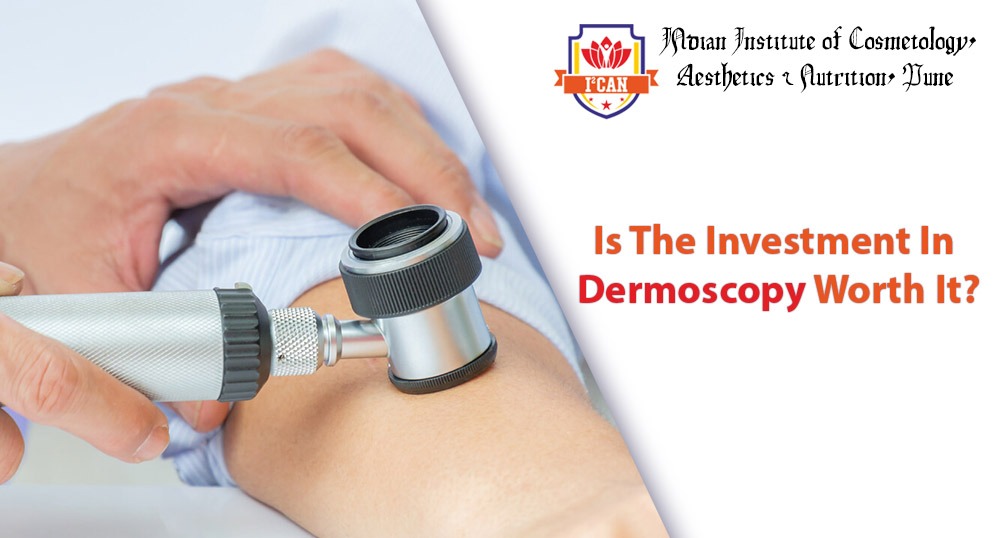
Friedman created the word “dermoscopy,” which is also called dermatoscopy, surface microscopy, or epiluminescence microscopy. Dermoscope is a portable device with high resolution and is used to evaluate skin in a non invasive way. It is a relatively new, vibrant, and interesting field, aptly dubbed “The cosmetologist’s stethoscope.”It enables quick in vivo examination of the epidermis, dermoepidermal junction, and papillary dermis morphologic components which are not easily visible for a human eye.
Options Available For Dermatoscopes
1. Polarized dermoscopy Handheld
They have a magnification range of 10 to 20 times, don’t really need contact with skin or immersing fluid, and enable for speedy and secure inspection without risk of infection. Since these devices have the potential to distort colours, they have contact dermoscopy capabilities to reduce colour distortions.
2. Dermoscopy using nonpolarized light
The instrument emits a flash of light that strikes the skin at a 20-degree angle. In touch dermoscopy. A fluid such as oils, liquid, solution, glycerol, or methanol gel is applied between the skin and the instrument to minimize distortions and hence improved visualisation. These gadgets are not recommended since they have the potential to impair vessel visibility (due to pressure) and/or scaling (when employing a liquid interface).
3. Dermoscopes with video
These devices require a USB connection to be linked to a watching screen. The benefits include a wider view that can be shared with patients/ dermatologists and the ability to store photographs. Video dermoscopes have become quite affordable and these are usually the devices of choice for those who are starting in the field.
4. UV dermoscopy and multispectral dermoscopy
Surface pigmentation is better seen under blue light, superficial vascularity is better seen under yellow light, and deeper pigmentation and vascularity is best seen with red light, which reaches deeply.
Dermoscope’s Applications
Dermoscopes have multiple utilities, some of which include:
1. Dermoscopy for tinted and non-tinted skin cancers, such as melanocytic and non-melanocytic skin tumours, as well as benign and malignant skin tumours.
- Dermoscopy gives helpful and trustworthy information on the histopathologic subtype of basal cell carcinoma (BCC) and shows tumour features like ulceration or pigmentation, allowing for more appropriate tumour therapy.
- Dermoscopy allows for a more precise assessment of the true extent of the tumour, allowing for a more precise estimation of the required surgical margins and thereby lowering recurrence rates.
2. Inflammoscopy for psoriasis, lichen planus, pityriasis rosea, and other inflammatory illnesses. The treatment options of papulosquamous disorders become easier due to specific functionalities on dermoscopy, allowing for the recognition of dermatitis (yellow crusts and patchy dotted vessels), lichen planus (Wickham striae), and infections of the skin (peripheral white scales).
3. Entomodermoscopy for parasitic, fungal, respiratory, bacterial, or plasmodium infections that induce skin problems and invasions. Dermoscopy can be used to identify both frequent and unusual pest infestation, with scabies being an excellent example exhibiting the delta-wing jet with contrail.
4. Trichoscopy, which is used to diagnose hair and skin problems
Rudnicka and Olszewska invented the name in 2006. Trichoscopy increases diagnostic performance and may aid in a greater understanding of hair condition pathophysiology.
5. Abnormalities of the nail/nail fold
- It can be used to diagnose tiny, asymptomatic, or subungual tumours, as well as to check the response to therapy after surgery. Sores that respond to therapy indicate a reduction in capillary density inside viral warts.
- Dermatoscopy aids in the detection of cancers and the visualisation of vascular patterns. It makes defining surgical margins easier before therapy.
6. Various diseases
A yellow-orange backdrop is used as a dermoscopic clue to diagnosing systemic inflammatory skin diseases such as tuberculosis and eczema lupus.
7. During observation, dermoscopy is used
- Dermoscopy has been shown to be useful in the therapeutic monitoring of imiquimod treatment.
- Vitiligo activity has been assessed using dermatoscopy. Balakrishna et al employed a dermatoscopy to track the outcome of different vitiligo surgical operations.
Main Considerations While Using Dermoscopy
1. Vascular anatomy/ morphology and organisation
2. Colours
3. Patterns of growth
4. Deformities of the follicles
5. Characteristics
Benefits of Dermoscopy
- It improves diagnosis accuracy by reducing the ambiguity of clinical examination.
- It allows cancers to be detected at an earlier stage, improving the prognosis.
- It eliminates the necessity for biopsies that aren’t necessary.
- It gives recommendations for the best biopsy site, boosting histopathological accuracy.
Final Thoughts
Dermoscopy is an indispensable, transparent, and useful supplementary tool to assist in the recognition/differentiation of various skin disorders, representing their histological foundation, and so having a significant impact on dermatology clinical practice. As a result, it is well worth the expense.